New microscope enables deep and wide neuroimaging
GA, UNITED STATES, November 6, 2024 /EINPresswire.com/ -- This research introduces new techniques to significantly improve the field of view of three-photon microscopy. Scientists can now observe the activity of large groups of neurons in deep brain regions with unprecedented detail. This advancement enables a deeper understanding of brain function and neural networks.
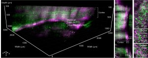
Researchers at Cornell have unveiled an advanced imaging technology capable of unprecedented deep and wide-field visualization of brain activity at single-cell resolution. The innovative microscope, named DEEPscope, combines two-photon and three-photon microscopy techniques to capture large-scale neural activity and structural details that were previously inaccessible.
Traditional multiphoton microscopy, a cornerstone for deep-tissue imaging, faces significant limitations in imaging depth and field of view, especially in highly scattering biological tissues like the brain. To prevent thermal damage, the imaging depth is usually increased at the expense of an exponentially shrinking field of view, making it challenging to observe large-scale neuronal networks. DEEPscope addresses these constraints by integrating a suite of novel techniques, allowing researchers to visualize extensive brain regions at unprecedented depths.
Key to this advancement is DEEPscope's adaptive excitation system and multi-focus polygon scanning scheme, which enable efficient fluorescence generation for large field-of-view imaging. These innovations make it possible to achieve high-resolution imaging across a 3.23 x 3.23-mm2 field with imaging speed that is sufficient for capturing neuronal activity in the deepest cortical layers of mouse brains. The ability to perform simultaneous two-photon and three-photon imaging further enhances the system’s versatility, allowing detailed exploration of both shallow and deep regions.
In their study, the researchers demonstrated DEEPscope's capability to image entire cortical columns and subcortical structures with single-cell resolution. They successfully recorded neuronal activities in deep brain regions of transgenic mice, observing over 4,500 neurons in both shallow and deep cortical layers. Moreover, DEEPscope enabled whole-brain imaging of adult zebrafish, capturing structural details at depths greater than 1 mm and across a field wider than 3 mm—a first in the field of neuroscience.
“DEEPscope represents a significant advancement in brain imaging technology,” said Aaron Mok, the study's lead author. “For the first time, we can visualize complex neural circuits in living animals at such a large scale and depth, providing insights into brain function and potentially opening new avenues for neurological research.”
The demonstrated techniques can be readily integrated into existing multiphoton microscopes, making it accessible for widespread use in neuroscience and other fields requiring deep-tissue imaging. By overcoming previous limitations, DEEPscope sets a new standard for large-field, high-resolution, deep imaging of living tissues, promising to advance our understanding of the brain’s intricate networks and their role in health and disease.
DOI
10.1186/s43593-024-00076-4
Original Source URL
https://doi.org/10.1186/s43593-024-00076-4
Funding information
This work was supported by the National Science Foundation NeuroNex (grant no. DBI-1707312), NIH/NINDS (grant no. U01NS103516), and Cornell Neurotech Mong Fellowship.
Lucy Wang
BioDesign Research
email us here
Legal Disclaimer:
EIN Presswire provides this news content "as is" without warranty of any kind. We do not accept any responsibility or liability for the accuracy, content, images, videos, licenses, completeness, legality, or reliability of the information contained in this article. If you have any complaints or copyright issues related to this article, kindly contact the author above.


