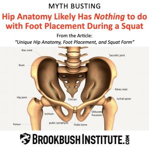
Busting Myths with Research: Your "Hip Anatomy" is not unique, and it does not affect "Squat Foot Placement."
The Brookbush Institute explains the research on hip morphology, squat foot placement, and makes a better recommendation for improving squat form.
Squat Stance Myth Busting:
Modifying foot position or stance width during a barbell squat to compensate for "unique hip anatomy" is actually a pretty weird idea. Most individuals have similar, or at least proportional bone structures. It seems to be a logical leap to jump from the hip joint to addressing foot positioning (skipping the knee). And, what happened to addressing mobility (e.g. reduced knee extension, hip flexion, and/or ankle dorsiflexion), muscle activation (e.g. gluteus medius activity, tibialis anterior activity, etc.), and/or strength issues of the primary muscles worked (e.g. gluteus maximus, quadriceps, and soleus strength). It is unclear why any personal trainer, strength coach, or clinical professional would want to jump to conclusions regarding hip morphology and squat form. And, this concept has implications for most lower-extremity strength exercises, including back-loaded squat foot placement, low bar squat foot placement, leg press foot placement, hack squat variations, smith machine squat, sumo squats, leg press, etc. Once you have had a chance to read the full article, we think you might agree that the foot placement angle for most lower extremity exercises should be toes forward, feet hip to shoulder width, and that discomfort in this position is not evidence of structural issues, is not serious, but may be a sign that optimizing performance will include additional mobility or corrective exercise techniques.
Hip Anatomy and Foot Position Summary:
- Normally Distributed: Research suggests that variations in hip anatomy are normally distributed, and could be plotted on a "bell curve”. That is, the gross majority of individuals exhibit bone shape, angles, and alignment, that are within a relatively small range of variation.
- Little to No Correlation: There may be no correlation between hip morphology and foot placement. In fact, it is likely that other structural angles compensate for normal variations in hip morphology during development.
- A Logical Error: Excessive hip retroversion and excessive hip anteversion cannot both be addressed by the same recommendation (feet wider and/or feet turned out). Further, if the standard deviation in hip anteversion is about 10°, how does anyone justify 20 - 50° of feet turn out during squats?
- A Functional Anatomy Error: Feet turn out is not hip external rotation, it is tibia (knee) external rotation.
- Correlated with Pain, Dysfunction, and Injury: Research demonstrates that feet turn out and knees wide (functional varus) are correlated with pain, dysfunction, and/or a higher risk of injury.
- A Better Solution: Before accepting small imperfections in movement as evidence of permanent, life-long, structural abnormalities, it may be recommended that these very addressable and common issues (e.g. feet turn out, knees bow in, etc.) are targeted with a "corrective exercise" or "movement prep" routine. Research has demonstrated that corrective exercise can improve alignment, and performance and reduce the risk of injury.
- Our Recommendation is Simple. Perform a movement assessment (like the Overhead Squat Assessment), address the issues you identify with corrective exercise, and then modify squat form, if necessary, based on comfort or performance needs. Chances are that addressing issues with corrective exercise noted during a movement assessment will greatly reduce the amount of compensation necessary to feel comfortable during a squat.
For the complete article and a sample movement preparation program, check out: Squat From, Hip Anatomy, and Foot Placement
Selected Citations (Full annotated bibliography included in the article linked above)
Hip Morphology:
2. Eckhoff, D. G., Kramer, R. C., Watkins, J. J., Alongi, C. A., & Van Gerven, D. P. (1994). Variation in femoral anteversion. Clinical Anatomy: The Official Journal of the American Association of Clinical Anatomists and the British Association of Clinical Anatomists, 7(2), 72-75.
5. Sengodan, V. C., Sinmayanantham, E., & Kumar, J. S. (2017). Anthropometric analysis of the hip joint in South Indian population using computed tomography. Indian journal of orthopaedics, 51, 155-161.
6. Saikia, K, Bhuyan, S, and Rongphar, R. Anthropometric study of the hip joint in Northeastern region population with computed tomography scan. Indian Journal of Orthopaedics 42(3): 260, 2008.
10. Pierrepont, J. W., Marel, E., Baré, J. V., Walter, L. R., Stambouzou, C. Z., Solomon, M. I., ... & Shimmin, A. J. (2020). Variation in femoral anteversion in patients requiring total hip replacement. HIP International, 30(3), 281-287.
Feet Out Correlated with Dysfunction:
11. Willson, J. D., & Davis, I. S. (2008). Lower extremity mechanics of females with and without patellofemoral pain across activities with progressively greater task demands. Clinical biomechanics, 23(2), 203-211.
12. Winslow, J., & Yoder, E. (1995). Patellofemoral pain in female ballet dancers: correlation with iliotibial band tightness and tibial external rotation. Journal of Orthopaedic & Sports Physical Therapy, 22(1), 18-21.
13. Lo, G. H., Harvey, W. F., & McAlindon, T. E. (2012). Associations of varus thrust and alignment with pain in knee osteoarthritis. Arthritis & Rheumatism, 64(7), 2252-2259.
14. Skou, S. T., Wrigley, T. V., Metcalf, B. R., Hinman, R. S., & Bennell, K. L. (2014). Association of knee confidence with pain, knee instability, muscle strength, and dynamic varus–valgus joint motion in knee osteoarthritis. Arthritis care & research, 66(5), 695-701.
Corrective Exercise Improves Performance
17. Bennell, K. L., Dobson, F., Roos, E. M., Skou, S. T., Hodges, P., Wrigley, T. V., ... & Hinman, R. S. (2015). Influence of biomechanical characteristics on pain and function outcomes from exercise in medial knee osteoarthritis and varus malalignment: exploratory analyses from a randomized controlled trial. Arthritis care & research, 67(9), 1281-1288.
18. Bell, D. R., Oates, D. C., Clark, M. A., & Padua, D. A. (2013). Two-and 3-dimensional knee valgus are reduced after an exercise intervention in young adults with demonstrable valgus during squatting. Journal of athletic training, 48(4), 442-449.
19. Crow, J. F., Buttifant, D., Kearny, S. G., & Hrysomallis, C. (2012). Low-load exercises targeting the gluteal muscle group acutely enhance explosive power output in elite athletes. The Journal of Strength & Conditioning Research, 26(2), 438-442.
Brent Brookbush
Brookbush Institute
Media@BrookbushInstitute.com
Visit us on social media:
Facebook
Twitter
LinkedIn
Instagram
YouTube
TikTok
Legal Disclaimer:
EIN Presswire provides this news content "as is" without warranty of any kind. We do not accept any responsibility or liability for the accuracy, content, images, videos, licenses, completeness, legality, or reliability of the information contained in this article. If you have any complaints or copyright issues related to this article, kindly contact the author above.

