Improving resolution and reducing noise in fluorescence microscopy with ensured fidelity
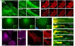
USA, August 7, 2024 /EINPresswire.com/ -- Scientists developed a new method to improve resolution and reduce noise in fluorescence microscopy images. This technique utilizes mathematical tools to analyze and enhance image details, specifically addressing the fidelity of computational resolution extension. It outperforms existing methods by boosting signal-to-noise ratio and achieving higher resolution without introducing artifacts. This paves the way for more precise and informative imaging in various microscopy applications.
Fluorescence microscopy is a cornerstone of modern biological imaging, allowing scientists to study cells and their processes in real time. However, limitations in resolution and noise levels can hinder the clarity and detail of these images. Moreover, the laser illumination could cast toxicity on cells and cause photo-bleaching, further confining the signal-to-noise ratio (SNR) in live-cell imaging. Researchers have developed a new deconvolution method that addresses these challenges, significantly improving image quality without introducing artifacts. This simultaneously brings new possibilities for live-cell imaging.
This novel technique renews both the noise-control model and the resolution extension mechanism in the deconvolution method. It utilizes a mathematical framework called multi-resolution analysis (MRA) to analyze fluorescence microscopy images for noise control. The method capitalizes on two critical characteristics of fluorescence images: sharp contrast across edges and smooth continuity along those edges, originating from the physical properties of excited fluorophores. Researchers have shown that this approach can distinguish useful signals from noise more precisely than previously mainstream variation-based methods.
The researchers reflect the deficits of previous mainstream statistical Richardson-Lucy iteration, which tends to produce artifacts rather than real high-frequency information. They find an alternative approach by incorporating the proposed edge-driven noise control method into a model-solution framework. An acceleration approach is also proposed to allow sufficient iterations in a short computation time. As a result, MRA can improve the SNR and resolution of fluorescence images with better noise resistance and ensure fidelity. The MRA deconvolved results can be verified by physical super-resolution microscopes, even if the structure is very complex, outperforming previous statistical deconvolution methods.
To manage situations with heavy background noise, the researchers further devise SecMRA, incorporating a bias thresholding mechanism for computational sectioning. It performs better than conventional methods and allows scientists to perform more challenging imaging tasks with severe background noise or low SNR.
This breakthrough has significant implications for various fluorescence microscopy applications. For instance, researchers can now discern features as small as sixty nanometers using structured illumination microscopy (SIM) in a fidelity-ensured manner, a feat previously limited by resolution constraints. Additionally, SecMRA facilitates the long-term observation of interactions between cellular structures, providing valuable insights into cellular processes.
The research team behind this innovation emphasizes the importance of maintaining fidelity in deconvolution. Unlike some existing statistical methods that can introduce artifacts, the MRA approach ensures the accuracy and reliability of the enhanced images. The parameters in the MRA pipeline do not significantly influence the obtainable high-frequency information, boosting the objectivity in the deconvolution process.
The authors have also proposed a solution for adjusting hyperparameters in the regularized deconvolution algorithm to promote this technology. They achieved automatic parameter determination by estimating the noise strength using the sparsity of the curvelet coefficients. At the same time, to facilitate fellow researchers, the authors have open-sourced all the source code and the original fluorescence image data and written interactive GUI software to facilitate users' and developers' use. The authors hope that MRA can become a new generation of high-fidelity deconvolution tools widely used by biologists and microscopists.
Moreover, for the thriving fields of computational super-resolution, sparse deconvolution, and deep learning, the authors hope that the physical truth judgment proposed in this work can become a widely used tool for authenticity judgment, and by open-sourcing related low- to high-resolution physical imaging datasets, provide an objective evaluation standard to further enhance the performance of different algorithms.
DOI
10.1186/s43593-024-00073-7
Original Source URL
https://doi.org/10.1186/s43593-024-00073-7
Funding information
This work was supported by the National Key R&D Program of China (2022YFC3401100) and the National Natural Science Foundation of China (62025501, 31971376, 92150301, 62335008).
Lucy Wang
BioDesign Research
email us here
1 https://doi.org/10.1186/s43593-024-00073-7

